Multiphoton Microscopy 
Multiphoton microscopy includes a variety of nonlinear optical techniques such as 2PF, 3PF, SHG, THG, CARS, SRS, etc. The key advantages of nonlinear methods is a tiny volume of light and matter interaction, which delivers a lower background noise, better spatial resolution, possibility of deep tissue as well as functional imaging.
Some nonlinear techniques are label-free, reduce the toxication of the tissue under observation and characterise the biochemical processes the closest to ones occurring in the living organisms. The high peak intensity of ultrashort laser pulses are required to maximize data acquisition rate and minimize tissue heating.
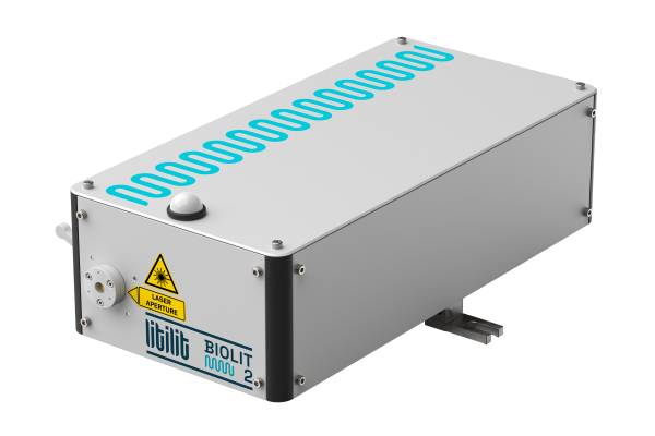
Biolit 2 is perfect choice for multiphoton microscopy. The laser delivers sub-80fs pulse duration with a nearly perfect Gaussian-like pulse shape and near diffraction limited beam quality. The lack of pulse pedestal and sideband satellites enables highly efficient signal generation in the tissue and suppression of background noise. The platform allows a wide range of pulse repetition rates from 15 MHz to 40 MHz.
The lower pulse repetition rate compared to common Ti:Sapphire oscillators provide higher peak power at the same avg. power level. This prevents thermal damage of the tissue. High peak to average power ratio facilitates up to 5x higher signal at the same average power compared to industry’s standard 80 MHz pulse repetition rate.
Biolit 2 has integrated tuneable dispersion pre-compensation (up to 10’000fs2) to make sure that maximal peak power and therefore nonlinear signal is achieved right on the sample, compensating for the dispersion of the imaging optics.
The compact laser head, passive air cooling and turn key operation simplifies the integration with a microscope. Using Biolit 2 laser you’ll get brighter and deeper images, faster.

Credits: Ludo van Haasterecht, Max Blokker, prof. dr. Marloes Groot, Vrije Universiteit Amsterdam, Frank van Mourik, Femto Diagnostics b.v.
The image of skin tissue, obtained with a multiphoton microscope using Biolit 2 femtosecond laser: red fibers – collagen bundles, blue lines – elastin fibers, green blobs – fat cells. Field of view – 500 micrometers.
Epilepsy brain tissue multi-contrast imaging
We are thankful to our customers from Vrije Universiteit Amsterdam for sharing extraordinary images of human brain tissue taken using different multi-contrast nonlinear imaging techniques with the help of Biolit 2. Normal appearing white matter was obtained from an epilepsy patient and a tissue block imaged over time while perfused with artificial cerebrospinal fluid. Multiphoton depth stacks were acquired with 3 channels: THG (green), SHG (red), and TPEF (blue). Colorful structures appear in this stack: some capillaries in red showing up in the first few micrometers. This is followed by myelinated axons (starting at z = 30 µm) in green.
Courtesy: Laura van Huizen, prof. dr. Marloes Groot, Vrije Universiteit Amsterdam, Geert Schenk, Antonio Luchicchi, Amsterdam UMC, Clinical Neuroscience.